Christiana Spine Center, PA is now offering Intracept by relievant. Please contact our office at 302-602-7000 to schedule a consultation today!
Please see video under Media tab for more details.
×

Services That We Provide
-
Minimally Invasive Spine Surgery

Minimally Invasive Spine Surgery (MISS) uses the latest advanced technology to treat a variety of spinal conditions. Special surgical instruments, devices and advanced imaging techniques are used to visualize and perform the surgery through small incisions. The aim of MISS is to minimize damage to the muscles and surrounding structures enabling faster recovery, less blood loss and lower chances of a post-operative infection. These advances have allowed us to perform many surgeries as outpatient or only an overnight stay in the hospital that otherwise would have required 3-4 days in the hospital.
Benefits of MISS
The benefits of MISS over the traditional open spine surgery include:
- Smaller incisions
- Less risk of infection
- Minimal blood loss during the surgery
- Minimal post-operative pain
- Quicker recovery
- Shortens the hospital stay
- Quicker return to work and normal activities
Conditions suitable for MISS
Some of the spinal conditions treated using MISS techniques are:
- Degenerative disc disease
- Scoliosis
- Spinal stenosis
- Spondylolisthesis
- Fractures
- Herniated discs
Procedure
Minimally invasive spine surgery is performed through smaller skin incisions. While the skin incisions are smaller, the true benefit of minimally invasive surgery is the decreased injury to the deep muscles of the spine. Drs. Fisher and Murray are experts in minimally invasive techniques allowing a faster recovery and less pain while still accomplishing all of the goals of traditional open surgery. Our surgeons utilize surgical microscopes and techniques including minimally invasive lateral (from the side) lumbar surgery and computer assisted guidance. Computer guidance allows us to place spinal implants with accuracy of less than 1 milimeter with making an incision just large enough to place the implant. This allows for unparalleled safety and the least invasive technique possible.
Risk and Complications
As with any surgical procedure, there are risks involved with minimally invasive spine surgery. The risks and complications of MISS may include infection, bleeding, nerve injury, as well as complications due to general anesthesia.
- Meet the Surgical Team
- Surgical Procedures
- Pre and Post-Operative Instructions
- Patient Forms
Meet the Surgical Team
Please click on the below tabs to know more about Surgical Procedures
Lumbar Microdiscectomy
Lumbar Laminectomy
Disc Replacement Surgery (cervical (neck) and lumbar (back))
Anterior Cervical Discectomy and Fusion (ACDF)
Minimally Invasive Lateral Lumbar Fusions (XLIF, DLIF)
Anterior Lumbar Interbody Fusion (ALIF)
Posterior Cervical Fusions and Laminoplasty
Pre and Post-Operative Instructions
- Surgery Date: _____________________ Time: ________________ Arrive: ____________________
- Patients may have NOTHING TO EAT after MIDNIGHT prior to surgery. If you eat anything after midnight, your surgery will be cancelled.
- You are allowed to drink WATER OR GATORADE ONLY 4 hours prior to surgery and 2 hours prior to arrival. If you have any questions, please call your doctor.
- Arrive to the hospital two hours prior to your scheduled surgery. You will need to go to the first floor - Surgery and Procedure Unit. If you are not sure where to go, check with the receptionist in the main lobby of the hospital.
- You will need to be seen six weeks after surgery. Your appointment is scheduled for ______________________ at ___________________. It is very important that you keep your six week appointment. Due to limited office hours, rescheduling this appointment can be difficult.
Patient Forms
You will need the Adobe Reader to view and print these documents.

Non-Surgical Treatment
Our physicians at Christiana Spine Center are skilled in offering many conservative treatments to treat specific spinal problems without any surgical intervention. Spinal injections can be administered to relieve pain and symptoms, while physical medicine and rehabilitation, in the form of exercise and massage, can be instituted to help ensure you recover completely.
- Meet the Non-Surgical Team
- Physical Medicine & Rehabilitation
- Radiofrequency Ablation Information
- Spinal Injections
Meet the Non-Surgical Team

Tony R Cucuzzella, M. D.
Physical Medicine & Rehabilitation
View profile
Fluoroscopic Spine Procedures


Scott T. Roberts, M. D.
Physical Medicine & Rehabilitation
View profile
Fluoroscopic Spine Procedures
Physical Medicine & Rehabilitation
Physical Medicine and Rehabilitation (PM&R) also called physiatry, is a branch of medicine concerned with diagnosing and treating disorders and injuries that involve movement. The physicians are known as physiatrists who are experts in diagnosing and treating pain (acute or chronic). The physiatrists help patients to achieve optimal quality of life and function by reducing pain. This branch of medicine comprises of various treatment options, including interventional pain management such as facet injections, spinal cord stimulation and epidural injections. Interventions in physical therapy may include massage, braces and exercise programs.
Epidural Steroid Injections
Steroids are chemicals that are naturally present in our body. Synthetic steroids are generally used in the treatment of inflammatory conditions such as spinal disc injury and degenerative diseases among others.
Facet Injections
Facet joint injections contain a strong anti-inflammatory agent called corticosteroid and an anesthetic for pain relief. They are given to relieve pain in the back, neck, arm and leg and even headaches caused from inflammation of the facet joints.
Nerve Block Injections
A selective nerve block is the injection of an anesthetic and steroid medication around the spinal nerve root to diagnose or treat pain. It is indicated to relieve pain, weakness, numbness and tingling sensation in your neck, back.
Sacroiliac (SI) Joint Injections
The sacroiliac joint connects the lowest part of the spine, the sacrum, to the adjoining bones of the pelvis, the iliac bones that are present on either side of the sacrum. In an adult there is minimal movement at these joints.
Radiofrequency Ablation
Radiofrequency ablation (RFA) also called rhizotomy or neurotomy is a novel non-surgical technique of treating pain. This technique employs radiofrequency waves to produce heat and the heat produced damage the nerves transmitting pain signal to the brain.
Spinal Cord Stimulation
Spinal cord stimulation (SCS) is used for the management of chronic pain in arms and legs that has not responded to conventional modalities of treatment. Specific segments of the spinal cord are stimulated through electrical signals.
Spinal Bracing
Spinal braces are external devices that help in the management of spinal disorders. They restrict the movement of the affected region of the spine, as well as support and stabilize it to relieve pain and promote healing.
Exercise Programs
Exercise is the most significant way to improve the health and decrease pain. Appropriate exercises can increase strength, improve flexibility, and reduce back pain. Most of the doctors accept that exercise plays.
Physical Therapy
Physical therapy is a health care specialty concerned with diagnosing, evaluating, and treating disorders and injuries of musculoskeletal system. The purpose of physical therapy is to help each patient restore their maximal level.
Radiofrequency Ablation
Radiofrequency ablation (RFA) also called rhizotomy or neurotomy is a novel non-surgical technique of treating pain. This technique employs radiofrequency waves to produce heat and the heat produced damage the nerves transmitting pain signal to the brain. This procedure is performed to treat painful facet joints in the spine that usually cause chronic low back pain and neck pain.
Radiofrequency ablation treatment is considered only after it is confirmed that the cause of back pain lies in the facet joints and this is confirmed by performing a diagnostic facet joint injection. Facet joint injection relieves pain for a short duration whereas radiofrequency ablation can keep you pain-free for a longer period of time.
Radiofrequency ablation is a minimally invasive technique and therefore administration of general anesthetic is not required. You will be conscious throughout the procedure and lying on your stomach. Only a small area over your back which requires treatment is cleansed and numbed. This procedure is performed under the guidance of fluoroscopy. The fluoroscope is a special kind of X-ray machine that helps doctors to visualize the placement of the needle electrode in invasive procedures.
During the procedure, your doctor will direct a special radiofrequency needle electrode close to the facet joint in such a manner that the needle tip lies almost near to the medial branch nerve. The needle tip is then heated so that the nerve gets cauterized and destroyed thereby reducing the pain. This procedure may last for about an hour or two.
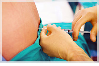
Spinal Injections
Patient Information for Spinal Injections at the Christiana Spine ASC
What can I expect during a spinal injection?
Only the patient is allowed in the procedure room during the injection. We ask that the patient eat a light meal, drink and take their normal medications. During most procedures, an IV needle will be placed in your hand or arm in case any medication is needed during the procedure. Blood pressure and heart rate are monitored. The procedure can take anywhere from 5-30 minutes. NUMBING MEDICATION IS USED LIBERALLY AND IN MOST CASES THE PROCEDURE IS NOT SIGNIFICANTLY PAINFUL. A small amount of dye will be injected to help with visualization during the procedure so please let us know if you have an allergy to x-ray dye or contrast dye. You should plan on having a ride home.
Why should I have a spinal injection?
The purpose of the injection is to carry medication to the inner part of the spine where the more serious types of strain and injury can affect the disc and spinal joints, causing pressure or irritation of the nerves, or pain arising from the joints. In most cases, the medication that is going to be used is a very potent anti-inflammatory steroid. Many doctors and patients refer to this medication as "cortisone", although hydrocortisone is rarely used anymore. There are better types of medication similar to hydrocortisone. In this office, we usually use triamcinolone or dexamethasone, which are in the same family of medications as hydrocortisone. The goal of the injection is to deliver this potent anti-inflammatory steroid as close as possible to the anatomical structure that we believe is causing your pain, thus providing the greatest chance of pain relief.
How long will it take before I see relief from the spinal injection?
The benefits from the injection may appear almost immediately or may build up gradually over seven to ten days. Increased pain may be experienced the day of the injection and the following day. Although unusual, this pain may last up to seven days.
How long will the benefits of the injection last?
Many patients ask how long the injection will last or if the injection "wears off". The medication injected is a potent anti-inflammatory and will cause a reduction in inflammation and pain in the area where it is injected. If the injection relieves 75% to 100% of your pain one (1) to two (2) weeks after the injection, most likely the results will last at least several months. It is highly recommended that a successful injection be combined with therapeutic stretching and strengthening exercises to prolong the benefits of an injection.
Are there any risks or complications from having this procedure?
Overall, these injections are very safe. Minor side effects are not uncommon, but severe complications are extremely rare.
Up to twenty percent (20%) patients experience dizziness, which may last for a few minutes during the procedure. This is generally due to a drop in blood pressure and resolves quickly. Intravenous fluids can be administered if this dizziness does not resolve quickly or is causing anxiety. Although unlikely, fainting may occur, and this would once again be due to a temporary drop in blood pressure. With cervical injections, loss of balance or unsteadiness may be present for 8 hours after the injection due to the anesthetic that is used.
Up to twenty percent (20%) of patients can experience numbness in the arms or legs after the procedure, and less frequently some patients may experience weakness in the arms or legs after the procedure. This sensation would generally last for one half hour but could last up to eight hours after the injection depending upon the type of anesthetic used during your procedure. For this reason, we would advise that you do not operate a vehicle or perform any activities that require coordination for twelve hours after the injection.
Minor or temporary reactions may result due to the use of corticosteroid medication (otherwise known as “cortisone” or “steroid” medication) with the procedure. Diabetic patients may experience a short-term increase in their blood sugars after the procedure (ranging from 1 day to 1 week), thus blood sugars should be checked at least daily for one week after your injection. Some patients may develop a headache or facial flush the following day, which lasts twelve to twenty-four hours. Rarely, some women can experience disruption of their menstrual cycle for one or two cycles.
Too many cortisone injections (or oral steroid preparations) over time can increase risk of osteoporosis (or bone thinning). The appropriate number of injections over times should be discussed with your physician.
Very rarely, a patient could develop an acute allergic reaction to the contrast dye that is used during the procedure. Medications will be available during the procedures that are effective at reversing allergic reactions. Less than one percent (1%) of patients will develop a headache, which can last up to twenty-four hours. Rest and contacting our office or your “family doctor” doctor is advised.
Other extremely rare complications have been reported, but their occurrence is so infrequent that an actual rate of occurrence is not available. These reported complications include infection and hip damage with possible need for an artificial hip.
Serious neurologic events, some resulting in death, have been reported with epidural injection of corticosteroids. Specific events reported include, but are not limited to, spinal cord infarction, paraplegia, quadriplegia, cortical blindness, and stroke. These serious neurologic events have been reported with and without use of fluoroscopy. The safety and effectiveness of epidural administration of corticosteroids have not been established, and corticosteroids are not approved for this use.
Corticosteroids (otherwise known as "cortisone" or "steroids"), such as dexamethasone, triamcinolone, methylprednisolone and betamethasone are not approved by the FDA for epidural injections. The use of corticosteroids for epidural injections is considered "off-label" use.
What if the injection does not work?
One possibility is that the medicine was not injected into the anatomic location that was the source of the pain. This means that your history and physical examination should be re-assessed to re-diagnose the problem. Although disappointing, an injection that does not provide pain relief often provides information to correctly diagnose your problem.
Another possibility is that the medicine was injected in the correct anatomic location, but that the problem causing your pain, such as a large slipped disc or severe spinal stenosis, is too great to be overcome by a simple injection. When this is the case, the patient will generally experience some very minor pain relief for several days and then the pain will return. When this occurs, it may not be worthwhile to perform any further injections.
What is the protocol for treatment after the first injection?
Often patients achieve pain relief that is lasting after just one injection. If indicated, a second or third injection may be performed to further any benefits that are gained from the first injection. Performing up to four (4) injections a year is considered safe and is a common protocol for many physicians who do these procedures. Further injections may or may not be worthwhile and will be determined on an individual basis at the follow up visit.
What do I need to do the day of the procedure?
Please bring a current list of all your medications with you along with your insurance information and picture ID. The ASC and office are two separate facilities. This will ensure your chart is the most up to date. As a courtesy to you, our office verifies benefits and obtains authorization with your insurance carrier prior to your visit at the ASC. It is your responsibility to provide facility co-payments and deductibles at the time of service. For any billing concerns, please call our Billing Office at (302) 602-7002 , between the hours of 8:00 A.M.-4:00 P.M. Monday through Friday. Lockers are provided to hold clothing and valuables. Do not wear jewelry. Bring a current list of all your medications with you. This list will ensure that our chart is the most up to date.
What should I expect on the day of the procedure?
Expect to be at the ASC for about 45-90 minutes. Times may vary based on type of procedure and if sedation is requested. If sedation is requested, no food or drink 2 hours prior to your procedure. If sedation is not requested, you can eat and drink normally. Lockers are provided to hold clothing and valuables. DO NOT wear jewelry.
For Patients Who Choose to Receive Sedation for Their Procedure
The Christiana Spine Ambulatory Surgery Center is pleased to provide the highest quality care during your Pain Management procedure.
In addition to local anesthesia, the facility offers a minimal sedation, Valium, which is optional. If preferred, you will receive this medication through an intravenous line (I.V.) that will cause you to relax.
Sedation will be specifically tailored to your individual needs on a moment-to-moment basis. Sedation is directed by a Board Certified Physician who will oversee the medical direction and supervision of your care. Medical devices will be utilized to carefully monitor your heart rate, blood pressure, oxygen saturation, and breathing.
You will then recover in the Post Anesthesia Care Unit (PACU) until you are fully ready for discharge. You will then be discharged from the facility with a responsible adult. You WILL NOT be permitted to receive sedation if you do not have a responsible adult and means of transportation other than yourself.
If you decide not to receive sedation prior to the procedure, and then you change your mind, you can let the staff know at the time of the injection and you will receive sedation as long as you have a responsible adult on the premises.
Everyone reacts differently to sedation; it may cause unsteadiness and drowsiness. You will be required to have a responsible adult remain with you throughout the day of your procedure. It is strongly advised that you DO NOT DRIVE for 24 hours after recieving sedation.
Possible side effects or complications associated with these procedures include:
Up to twenty percent (20%) patients experience dizziness, which may last for a few minutes during the procedure. This is generally due to a drop in blood pressure and resolves quickly. Intravenous fluids can be administered if this dizziness does not resolve quickly or is causing anxiety. Although unlikely, fainting may occur, and this would once again be due to a temporary drop in blood pressure. With cervical injections, loss of balance or unsteadiness may be present for 8 hours after the injection due to the anesthetic that is used.
Up to twenty percent (20%) of patients can experience numbness in the arms or legs after the procedure, and less frequently some patients may experience weakness in the arms or legs after the procedure. This sensation would generally last for one half hour but could last up to eight hours after the injection depending upon the type of anesthetic used during your procedure. For this reason, we would advise that you do not operate a vehicle or perform any activities that require coordination for twelve hours after the injection.
Minor or temporary reactions may result due to the use of corticosteroid medication (otherwise known as “cortisone” or “steroid” medication) with the procedure. Diabetic patients may experience a short-term increase in their blood sugars after the procedure (ranging from 1 day to 1 week), thus blood sugars should be checked at least daily for one week after your injection. Some patients may develop a headache or facial flush the following day, which lasts twelve to twenty-four hours. Rarely, some women can experience disruption of their menstrual cycle for one or two cycles.
Too many cortisone injections (or oral steroid preparations) over time can increase risk of osteoporosis (or bone thinning). The appropriate number of injections over times should be discussed with your physician.
Very rarely, a patient could develop an acute allergic reaction to the contrast dye that is used during the procedure. Medications will be available during the procedures that are effective at reversing allergic reactions. Less than one percent (1%) of patients will develop a headache, which can last up to twenty-four hours. Rest and contacting our office or your “family doctor” doctor is advised.
Other extremely rare complications have been reported, but their occurrence is so infrequent that an actual rate of occurrence is not available. These reported complications include infection and hip damage with possible need for an artificial hip.
Serious neurologic events, some resulting in death, have been reported with epidural injection of corticosteroids. Specific events reported include, but are not limited to, spinal cord infarction, paraplegia, quadriplegia, cortical blindness, and stroke. These serious neurologic events have been reported with and without use of fluoroscopy. The safety and effectiveness of epidural administration of corticosteroids have not been established, and corticosteroids are not approved for this use.
Corticosteroids (otherwise known as "cortisone" or "steroids"), such as dexamethasone, triamcinolone, methylprednisolone and betamethasone are not approved by the FDA for epidural injections. The use of corticosteroids for epidural injections is considered "off-label" use.
The benefits from the injection may appear almost immediately or may build up gradually over seven to ten days. Increased pain may be experienced the day of the injection and the following day. Although unusual, this pain may last up to seven days.
Minimal Sedation/Conscious Sedation – I understand that anesthesia in the form of minimal sedation (anxiolysis) may be needed so that my doctor can perform the procedure. It has been explained to me that all forms of anesthesia involve some risks. Generally minimal sedation is adequate, however, in some cases (either intended by the physician, or based on an individual's response to anesthetic medication), moderate sedation is achieved. Risks of sedation include depressed breathing requiring breathing tube, awareness under anesthesia, and injury to blood vessels. I understand that the type(s) of anesthesia to be used is determined by many factors including my health status, the type of surgery/procedure and my physician’s preference as well as my own desire. I consent to the anesthesia service deemed appropriate by the CSASC team.
Diagnostics: EMG & Ultrasound
Diagnosis of the exact source and cause of pain and disability is the key to choosing the most appropriate treatment, and achieving optimum outcomes of relief. Christiana Spine Center is well-equipped with the most up-to-date diagnostic tools to provide our physicians with the most accurate diagnosis of your particular condition.
Meet the Diagnostics Team
EMG
Your doctor may order an EMG test for you. The test can be done here at the Christiana Spine Center.
What is an EMG?
EMG stands for Electromyography. This is an electro diagnostic test that can study your nerves and muscles. You will lie on an examining table and a needle recording electrode will be placed into skeletal muscle. You may have some discomfort during needle placement. The electrode will be able to monitor the electrical activity of a skeletal muscle. The recordings will be displayed as electrical waveform. You will be asked to try to relax your muscles during the test and at certain times you may be asked to contract certain muscles.
Nerve conduction studies will also be included in the test. During this part of the test needles are not used. A small electrical current will be applied to various nerves at one site and the response is recorded at another site. This may cause a mild tingling feeling. The machine will be able to tell how fast the nerves conduct the current and if there are any problems with the nerves.
By examining the electrical activity your doctor will be able to tell if the muscles or nerves that go into these muscles are in any way abnormal. There is no lasting pain or side effects from having this test done.
What will an EMG be able to tell my doctor?
An EMG will be able to tell the doctor if there is any abnormal nerve function. This may help to find out the cause of your pain. The nerve conduction test tells how signals travel across the nerve. This helps to isolate the cause of abnormal function.
Is there any preparation for an EMG?
Inform your doctor if you are using a TENS unit, taking blood thinners, or have a cardiac pacemaker. Do not apply any lotion the day of the test. You may want to limit stimulants like coffee, tea, cola, and cigarettes for 2-3 hours before the test.
Ultrasound
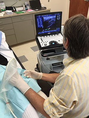
Medical ultrasound is a diagnostic medical imaging technique used to visualize muscles, tendons, ligaments, joints and many internal organs, to capture their size, structure and any pathological lesions with real time images. Ultrasound has been used by radiologists and ultrasound technologists to image the human body for at least 50 years and has become a widely used diagnostic tool. The technology is relatively inexpensive and portable, especially when compared with other techniques, such as magnetic resonance imaging (MRI) and computed tomography (CT). As currently applied in the medical field, properly performed ultrasound poses no known risks to the patient. Ultrasound does not use ionizing radiation such is with CT and X-Ray, and the power levels used for imaging are too low to cause adverse heating or pressure effects in tissue. Although the long-term effects due to ultrasound exposure at diagnostic intensity are still unknown, currently most doctors feel that the benefits to patients outweigh the risks. The ALARA (As Low As Reasonably Achievable) principle has been advocated for an ultrasound examination – that is, keeping the scanning time and power settings as low as possible but consistent with diagnostic imaging. Dr Delport’s ultrasound practice, which is accredited by the American Institute for Ultrasound in Medicine (AIUM), strictly follows the ALARA principle.
What are the common uses of ultrasound:
- Pain
- Swelling
- Infection
- Mass
- Ultrasound is also used to guide procedures such as injections and biopsies
How should you prepare
You should wear loose clothing, and you may be asked to change into a gown. Otherwise, no preparation is required prior to your ultrasound examination, and there are no contra-indications to having an ultrasound done. Patients with pacemakers and metal implants are safe to receive an ultrasound examination.
What does the equipment look like
The ultrasound machine consists of a computer console, video display screen and transducer or probe which is placed on your body during the scan. The transducer resembles a microphone and is attached to the ultrasound machine by a cord.
Some exams may use different transducers (with different capabilities) during a single exam. The transducer sends out inaudible, high—frequency sound waves into the body and then listens for the returning echoes from the tissues in the body. The principles are similar to sonar used by boats and submarines. The ultrasound image is immediately visible on a video display screen that looks like a computer or television monitor. A small amount of gel is put on the skin to allow the sound waves to best travel from the transducer to the examined area within the body and then back again.
What to expect during the procedure
For most ultrasound exams, you will be positioned lying face-up on an examination table that can be tilted or moved. Patients may be turned to either side or on occasion placed in a face down position.
After you are positioned on the examination table, the radiologist will apply a water-based gel to the area of the body being studied. The gel will help the transducer make secure contact with the body and eliminate air pockets between the transducer and the skin that can block the sound waves from passing into your body. The transducer is placed on the body and moved back and forth over the area of interest until the desired images are captured.
There is usually no discomfort from pressure as the transducer is pressed against the area being examined. However, if scanning is performed over an area of tenderness, you may feel pressure or minor pain from the transducer.
Once the imaging is complete, the clear ultrasound gel will be wiped off your skin. Any portions that are not wiped off will dry to a powder. The ultrasound gel does not stain or discolor clothing.
Most ultrasound examinations are completed within 30 minutes, although more extensive exams may take up to an hour.
After an ultrasound examination, you should be able to resume your normal activities immediately.
Is it important who performs your ultrasound examination?
Ultrasound requires extensive training in order perform and interpret an ultrasound study. Ultrasound is operator dependent, which means that the training and skill of the operator is essential to establish the correct diagnosis and to perform an appropriate, safe and effective procedure. Dr Delport has spent many years learning ultrasound through his radiology residency and musculoskeletal fellowship at Thomas Jefferson University Hospital. As mentioned before, Dr Delport’s ultrasound practice is the only practice in Delaware accredited by the American Institute for Ultrasound in Medicine. You are therefore guaranteed a high level of expertise. Dr Delport personally performs all the ultrasound examinations himself.
Discogram
What is a discogram?
A discogram is a diagnostic test that is performed to view and assess the internal structure of a disc and to determine which disc is the source of pain.
Why do I need a discogram?
A discogram helps determine the anatomical source of low back pain for the patient and enables the physician to view the disc itself. The results of the discogram may confirm the necessity for surgery and/or determine the exact cause of a person's back pain, which will increase the likelihood of a successful outcome.
What happens during the test?
The patient is awake during the test in order to tell the discographer what kind of pain is generated by the injection. The test is done as an outpatient procedure under sterile conditions. Medication will be given to help with relaxation. The patient will be positioned on their abdomen, their lower back will be cleaned with an antiseptic and the doctor will insert a needle with a local anesthetic into the skin to numb the area. An X-ray machine (fluoroscopy) is used to help guide the needle into the suspected problematic disc and then a radiopaque dye is injected through the spinal needle into the center of the disc. A patient's pain may be replicated due to the pressure created by the dye. A CT is performed after the dye is injected to obtain images of the dye distribution. You will be discharged 1-2 hrs after the procedure.
Following the discogram...
Arrange to have a ride home. Drink plenty of fluids to clear dye from the body. Expect to have localized pain and increased discomfort for 2-4 days after the procedure.
Considerations
Prior to the injection, please notify us if there is any allergy to contrast dye or if you are pregnant. The incidence of disc infection is rare and is reported at 7 per 1000 per disc level tested. Nevertheless, antibiotics are injected into the disc to decrease the risk.
How do I get ready?
Do not eat solid foods after midnight the day before the test. Dress comfortably on the day of the test and try to relax.


-

Christiana Spine Center
Medical Arts Pavilion II
4735 Ogletown-Stanton Road,
Suite 3302
Newark, Delaware 19713.1101 Twin-C Lane,
Suite 202
Newark, Delaware 19713. -

Ambulatory Surgery Center
1101 Twin-C Lane,
Suite 102
Newark, Delaware 19713.Phone: (302) 602-7000
Infectious Control Line: (302) 996-9525
Fax: (302) 602-7012
View Details